
AI in medical imaging is about capacity, quality, and time-to-result. Scan volumes continue to rise while staffing lags, resulting in increased scheduling, reading, and follow-up demands. In many regions, radiologists’ workloads have nearly doubled, creating backlogs that hit service levels and revenue.
When AI is integrated into everyday tools, such as PACS (Picture Archiving and Communication System), EHR (Electronic Health Record), and RIS (Radiology Information System), the gains are measurable: faster turnaround times on critical cases, more consistent follow-ups, and fewer re-reads.
Demand is real: per-capita CT usage is up ~57% and MRI usage is up ~80% since 2007. Supply is tight: the UK reports a ~29% shortfall in consultant radiologists, and U.S. workloads have nearly doubled. In this context, structured reporting and assisted contouring reduce effort, while intelligent triage prioritizes urgent studies.
This article is intended for provider organizations, clinics, radiology departments, hospitals, imaging centers, teleradiology groups, medical device makers, radiology software vendors, and EHR developers. We focus on the strategic shift underway: from isolated algorithms to an operating model where artificial intelligence in medical imaging reliably reduces re-reads, accelerates routing, and standardizes reports without forcing clinicians to change tools.
If you’re planning the next imaging release or a platform roadmap, consider this your field guide to scaling AI, focused on data readiness, interoperability, security, privacy, and regulatory alignment end to end. And if you want a quick readiness check on data, integration, and governance, our healthcare IT consulting can help.
Highlights:
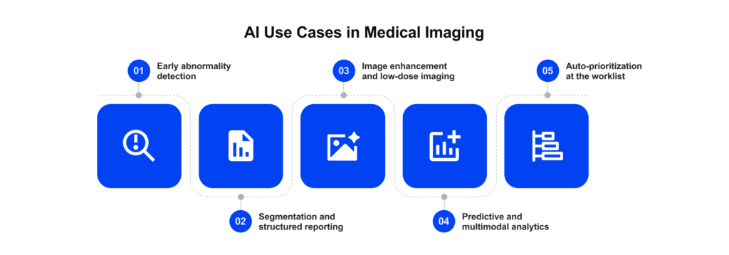
- AI-based medical imaging creates value today when it is integrated into PACS/EHR workflows.
- Early detection, triage, and QA shorten the time-to-read and reduce misses on high-volume CT and X-ray scans.
- Segmentation and structured reporting standardize follow-ups and speed multidisciplinary team prep.
- Image enhancement restores clarity from low-dose or shorter scans without changing radiologists’ tools.
- Real results depend on governed data, interoperability, security, privacy, and compliance by design.
What Can AI Actually Do in Imaging Today?
When AI in radiology is embedded into everyday workflows, its impact is measurable: faster reads on critical studies, more consistent follow-ups, and fewer re-reads. Below are the most proven capabilities, with real-world numbers.
Want these signals to roll up into decision-grade dashboards?
Explore our
healthcare data analytics servicesEarly abnormality detection
The most mature capability is early abnormality detection (production): models flag likely cancers, intracranial bleeds, pulmonary emboli, fractures, or suspicious nodules and surface cues directly in the DICOM viewer. This shortens the time-to-read for critical findings and reduces misses on high-volume modalities, such as CT, MRI, and X-ray.
The research shows that prioritizing chest X-rays (CXR) reduces report turnaround time (RTAT) for critical findings. For example, pneumothorax cases dropped from 80.1 to 35.6 minutes (p < 0.0001).
Segmentation and structured reporting
Segmentation means drawing precise boundaries around a region of interest in an image and separating it from the surrounding tissue. A radiologist or imaging engineer almost never works with the whole organ at once.
They focus on small areas and must distinguish very subtle changes in gray level. In CT or MRI, the segmented region becomes a 3D object that can be rotated, inspected from any angle, and measured consistently across studies.
For pre-surgical planning and tumor tracking, this precision is critical. Consistent volumes and margins make follow-up scans comparable, which speeds up MDT preparation. Multi-institutional head-and-neck CT auto-segmentation has shown 42-49% time savings.
Other studies report absolute savings from 14 to 93 minutes per case, depending on the anatomy. In practice, this means faster planning, standardized measurements, and less re-work, especially when the segmented region feeds directly into structured reports and downstream AI models.
Image enhancement and low-dose imaging
Image enhancing, denoising, deblurring, and super-resolution sit between pilot and production. Deployed at reconstruction or as a post-processing step, artificial intelligence in radiology recovers detail from low-dose CT or shorter MR sequences without disrupting radiologists’ tools. For instance, deep-learning reconstruction has shown a reduction of ~45% in effective dose across a large matched cohort.
Predictive and multimodal analytics
Predictive analytics derived from imaging patterns is typically conducted in a pilot/research setting, often integrated with EHR and lab data. These models estimate the risk of progression or complications to prioritize follow-ups and support tumor boards and care pathways.
Combining imaging with clinical data (labs, meds, problems) supports risk stratification and cohort prioritization for follow-ups and tumor boards; adoption is growing but remains pilot-heavy in most systems.
Need help with healthcare data & analytics?
We are here to help. Check our
Healthcare Data & Analytics ServicesAuto-prioritization at the worklist
Finally, auto-prioritization of high-risk studies is firmly in production, where PACS/RIS (Radiology Information System) integration is available: suspected hemorrhage, stroke, or major fracture cases jump the queue with alerts and timestamps.
With PACS and RIS integration, alerts push suspected hemorrhage, stroke, and major fractures to the top of the list. In CT pulmonary angiography, the report turnaround time (RTAT) decreased from 59.9 to 47.6 minutes, and the routine wait time decreased from 43.7 to 15.3 minutes.
For intracranial hemorrhage, flagged cases were initiated approximately 3.4 minutes sooner. Chest X-ray triage shows a reduction in RTAT of up to 77%. Large-vessel occlusion stroke programs report a ~44% faster arrival-to-diagnosis time.
Ready to surface results inside PACS/RIS/EHR across sites? See our healthcare integration solutions.
Read also:
Automating Prior Authorization with FHIR®How AI Fits into Imaging Workflows
Turning a model into something radiologists use every day requires clear roles between clinical and technical teams, reliable data flows, and stable links to imaging viewers, radiology information systems, and electronic health record systems.
This section explains how that usually works in practice. For example, consider a lung nodule screening program based on computed tomography scans. The same steps can be applied to other imaging pathways, such as magnetic resonance imaging (MRI) or ultrasound.
Data prepared for clinical use
Segmentation, annotations, and imaging QA sit mostly on the clinical side. Radiologists and imaging vendors define protocols, label structures, and agree on what counts as “ground truth.” Data and IT teams usually join once these rules and datasets exist. Their job is to standardize study metadata, link studies to patients and encounters, and turn informal rules into clear, queryable cohort definitions.
In a lung-nodule program, that means defining inclusion criteria, linking baseline and follow-up scans, and attaching outcomes so teams can study long-term performance and safety.
Pipelines for continuous learning and evaluation
On top of these curated datasets, teams build monitored pipelines with versioned data, models, and evaluation logic.
In the lung-nodule example, a pipeline can regularly re-score new cohorts, compare a candidate model to the current one, and run “shadow” evaluations before any change reaches the reading room. This helps catch performance drift when patient mix, scanners, or protocols shift over time.
Backends that embed AI into PACS/RIS/EHR
AI outputs are most useful when they are integrated into existing tools, rather than in a separate portal. Technical teams typically integrate results through DICOM and HL7® FHIR®, without touching source images.
For lung screening, a nodule category or recommended follow-up interval can be represented as a structured FHIR Observation or ServiceRequest linked to the patient and study, with the model version stored for traceability. Radiologists continue to use their usual viewer; the AI signal is simply an additional, auditable layer of context.
Clinician-facing views aligned to reporting
Product and radiology teams then shape worklists and summaries that match daily practice.
In the same pathway, a worklist might highlight patients due for follow-up, while a compact panel within the viewer displays interval growth, key measurements, and suggested next steps that can be integrated into a structured report. The goal is fewer clicks and clearer cues, not replacing clinical judgment.
Governance, version control, and infrastructure
Under the hood, mature setups track lineage for data, code, and models; capture detailed audit logs; and support safe rollbacks on containerized, GPU-ready infrastructure.
When a lung-nodule model is updated, each deployment can be tied to a specific dataset slice, evaluation report, approval decision, and timestamp. This end-to-end trail supports internal quality review, addresses regulator questions, and facilitates root-cause analysis in the event of an issue.
Before imaging results translate into decisions, leaders also need a view beyond DICOM: problems, medications, labs, encounters, and outcomes across sites and releases. FHIR-native data and analytics platforms, such as Kodjin, help bring together non-imaging context with imaging findings, allowing organizations to track the impact over time, compare cohorts, and see how changes in imaging AI actually affect care.

Build, Buy, or Partner? Choosing the Right Strategy for AI Medical Imaging Implementation
Getting AI for medical imaging is a trade-off across speed, scope, risk, and total cost. This guidance targets clinics and radiology departments, imaging centers, teleradiology groups, PACS/EHR vendors, and medical imaging AI startups. The table below compares the main paths and indicates when each is applicable.
| Buy pre-built | Build in-house | Partner (co-develop) | |
| Speed to value | Fast pilots and go-lives | Slow (hiring, data, infrastructure) | Moderate, faster with expert rails |
| Scope and fit | Fixed features, limited tuning | Tailored to your needs | Flexible: productize what matters |
| Integration | Vendor connectors, some gaps | Deepest, if you have bandwidth | Designed around your PACS/RIS/EHR |
| Regulatory evidence | Vendor documentation available, coverage may be uneven | You own all the evidence, SOPs, and traceability | Shared model, partner supplies templates/audit trails, you own final sign-off |
| Total cost of ownership (TCO) | Lower upfront, license growth | Higher upfront, lower opex later | Balanced; pay for acceleration |
| Talent & ops | Light internal ownership | Heavy: data, MLOps, SRE | Right-sized: you own, partner accelerates |
| Risk profile | Vendor lock-in, feature gaps | Delivery risk, team churn | Managed: roadmap control + expertise |
| Best for | Fast gap-filling, triage/QA | Strategic differentiation | Complex roadmaps, platform plays |
Now, let’s take a closer look at how each option plays out.
Buy pre-built AI solutions
Great for quick wins, triage, QA, and basic detection, when your priority is time-to-benefit. Expect fixed models and roadmaps, limited customization, and vendor-defined integration paths. Works best when you can live with “80% fit” and need results this quarter.
Build custom AI tools for medical imaginginternally
Best when AI is core to your differentiation, and you can fund data engineering, MLOps, validation, and clinician UX. You’ll get the tightest fit and full IP control, but you also own hiring, governance, and run costs. Timelines stretch without experienced platform teams.
Partner with an AI/healthtech provider
A middle path that keeps control of your roadmap while reducing delivery risk. You bring domain goals and ownership; your partner brings accelerators for data readiness, evaluation pipelines, secure integration, and compliance. The result: faster iterations, cleaner handoffs to PACS/RIS/EHR, and audit-ready deployments.
Edenlab helps imaging teams evaluate and design AI strategies across healthcare clients, from rapid vendor selections and PoCs to full platform builds.
We handle the complex aspects of operational AI: cleaning and mapping DICOM images and annotations, engineering continuous training/validation pipelines, integrating outputs into PACS/RIS/EHR via DICOM and HL7 FHIR, and developing clinician-first views. Our governance stack keeps releases compliant and reversible, so programs scale without re-platforming.
Recently, we partnered with Healthy.Mind to launch an AI-driven mental-health screening platform that detects ~80% of DSM-5 disorders in under 20 minutes. It’s clinically validated, tested with 1,500+ users, adapts to cultural context, and supports early intervention. As the team expands AI capabilities and readies a global rollout, the platform is also laying the groundwork for future agent-driven features that could reshape care delivery.
How Edenlab Helps with Medical Imaging and AI
AI in imaging only works on solid ground. Machine-learning models only work well when DICOM studies and annotations are complete and reliable, so teams start with data cleaning, label standardization, and clear rules for governance, version control, and secure access.
Integrations should follow how radiologists already work in their viewers, radiology systems, and electronic health records, instead of adding yet another standalone tool.
Security and privacy come built in. Healthcare risk is higher, so we embed compliance, access control, and protection at every layer. We work methodically and aim for real outcomes: clear KPIs like time-to-read, re-read rates, and throughput, with monitoring and rollback paths. Healthcare is the only domain we serve, so solutions move beyond demos and deliver impact in clinical and operational settings.
We recently partnered with a U.S. vendor to build an AI-powered analytics platform that helps clinical and research teams spot data-quality issues and explore patient cohorts across primary care and specialized domains like stem-cell research. Designed for AI from day one, it combines a graph-based backend, natural-language interaction, and a compliance-ready architecture, and it’s already informing the vendor’s next wave of agent-based tools.
Would you like to get similar results for your product? Lean on healthcare analytics product development.
Conclusion
Medical imaging with AI is already in use across real radiology services: triage that shortens time-to-read, segmentation that standardizes follow-ups, and quality checks that reduce re-reads. What separates a good demo from dependable value is the ecosystem around the model.
Successful deployments depend on a few essentials working in concert: a mature data and integration stack (DICOM/HL7 FHIR), smooth handoffs into PACS/EHR, clinician-aligned workflows and UX, and a compliance layer with version control, auditability, and rollback. With these in place, AI augments teams without added friction and remains maintainable as volumes, modalities, and regulations evolve.
Edenlab’s role is to make that ecosystem real. We prepare and govern the data, engineers repeatable pipelines, embed outputs where clinicians work, and implement the controls your risk and regulatory teams expect. The result is interoperability that delivers value today and a platform that can absorb tomorrow’s models without re-platforming or surprises.
AI won’t solve everything. But it can solve the right things reliably when it’s built on the rails that healthcare depends on. We’re here to help you put those rails in place.

Operationalize AI in radiology safely and at scale
We prepare datasets, engineer pipelines, and embed AI outputs into existing workflows to cut backlogs and reduce variability. Interoperability, privacy, and auditability included from day one.
FAQs
What AI use cases offer the fastest ROI in diagnostic imaging?
Triage and worklist prioritization, image-quality enhancement (low-dose CT/MR), and QA checks typically pay back first in the case of AI in diagnostic imaging. Structured reporting also assists by reducing re-reads and admin time. These use cases integrate quickly and show measurable time-to-read and throughput gains.
Can we implement AI without replacing our existing imaging software?
Yes. Most teams integrate through DICOM (including SR), HL7/FHIR bridges, or PACS/RIS plug-ins. We add event-driven services alongside your stack and stage rollouts with feature flags, eliminating the need for re-platforming.
How do we ensure regulatory compliance when using AI?
Treat compliance as an operating system: version everything, capture audit trails, and document intended use and performance. Align with FDA/MDR expectations, change control, cybersecurity, and post-market monitoring. Build evidence as you go, not at the end.
How much annotated imaging data do we need to train models?
It depends on the task and label type. Detection/triage often starts delivering with thousands of labeled studies; segmentation may need hundreds to low thousands of pixel-level masks. Transfer learning, curated cohorts, and ongoing hard-case mining reduce data demands over time.
What are the risks of using third-party AI tools in diagnostics?
Vendor lock-in, unclear provenance, bias and drift, and data-residency or security gaps. Mitigate with on-prem or VPC deployment options, documented model lineage, local validation against your cohorts, performance monitoring, and clear SLAs for updates and rollback.
Can AI reduce radiologist burnout?
Yes, when it cuts repetitive tasks and surfaces urgent cases first. Gains come from cleaner worklists, pre-filled structured reports, and fewer re-reads. Guardrails matter: good UX design and alert tuning prevent notification fatigue.
Stay in touch
Subscribe to get insights from FHIR experts, new case studies, articles and announcements
Great!
Our team we’ll be glad to share our expertise with you via email